


.png)

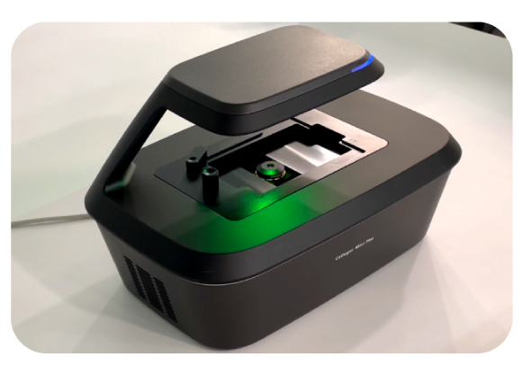
 Celloger® Mini Plus
- Automated live cell imaging system
- Celloger® Mini Plus is an automated live cell imaging system with advanced fluorescence and bright-field microscopy, autofocusing, and real-time multi-position imaging technology. It provides essential tools for acquiring high-quality images and obtaining accurate research results. This versatile and user-friendly system supports a wide range of cell-based research and applications. Expand your cell discoveries with Celloger® Mini Plus and accelerate your research.
Key Features
- Real-time cell monitoring
- Compact and portable design
- Multi-point imaging up to 96 wells
- Compatible with various vessel types
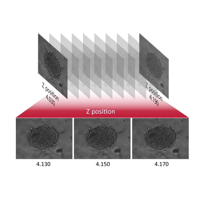
- Z-stack multi-focal imaging
- Reliable autofocus
Real-time monitoring
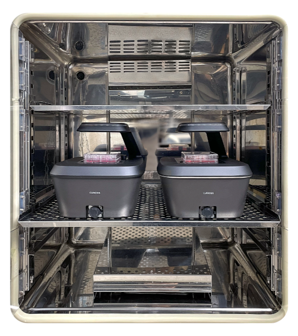
Maintaining the performance of a device working in a hot and humid environment is very challenging. With Celloger® Mini Plus, you can easily monitor live cells inside the incubator for a long time without disturbing the environment suitable for cell culture.
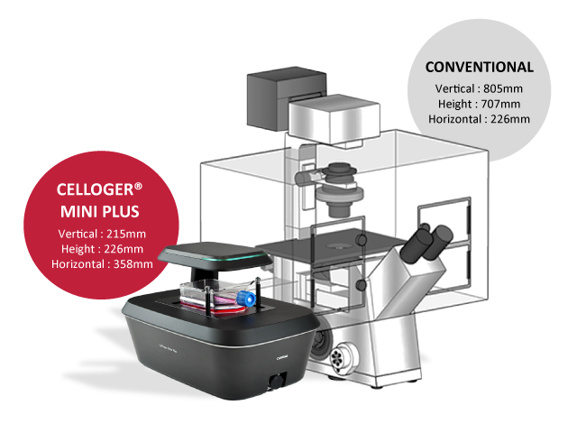
Compact size
Celloger® Mini Plus is compact in size with 226(h) x 358(l) x 215(w)mm where several Celloger® Mini Plus systems can fit into a standard CO2 incubator.
Since the system weighs about 5kg, it can be easily carried in and out of the incubator during the experiment.
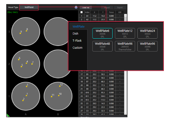
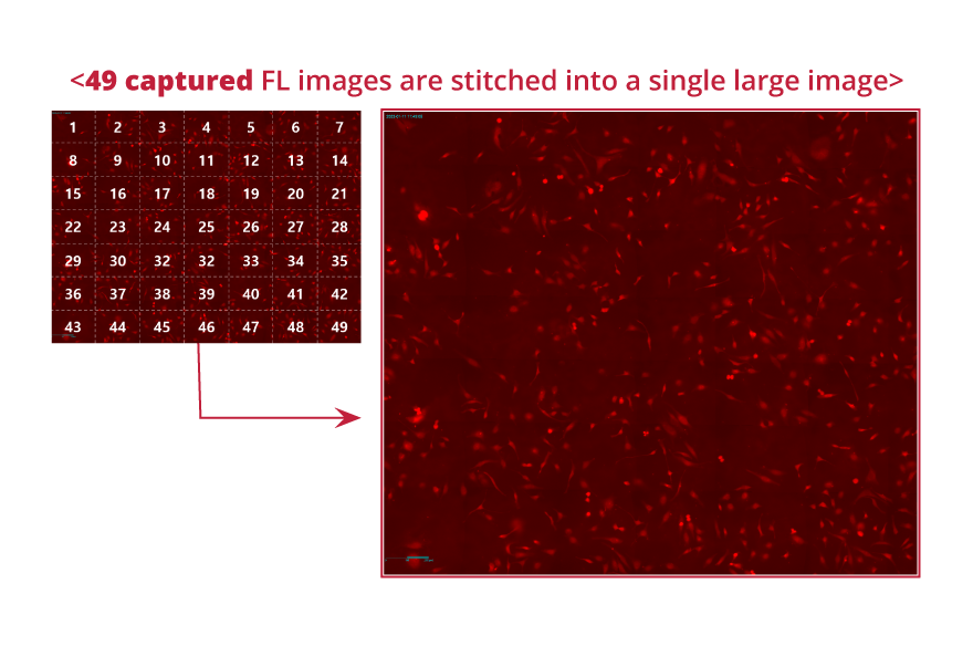
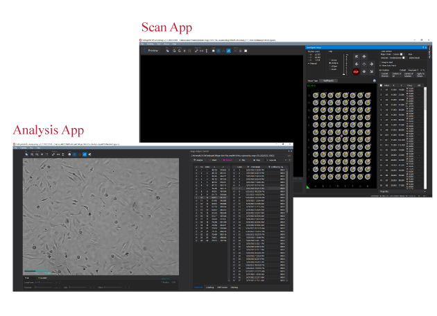
Multi-point imaging
Using the motorized camera that travels 117mm x 77mm, x and y axis respectively, multiple points within the travel range can be captured following the schedule (intervals, cycles, total time) set by the researcher.
Stable imaging performance
Celloger® Mini Plus doesn’t have a moveable stage but instead, the camera inside the system moves to capture the images of cell in multiple positions (refer to the video above). Since the vessel and cell sample are in a steady state, this provides a stable environment for the cells to grow.
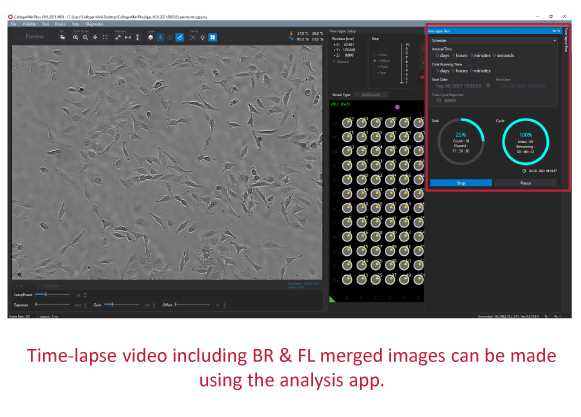
Time-lapse video
Images captured according to set schedule can be made into videos in simple clicks.
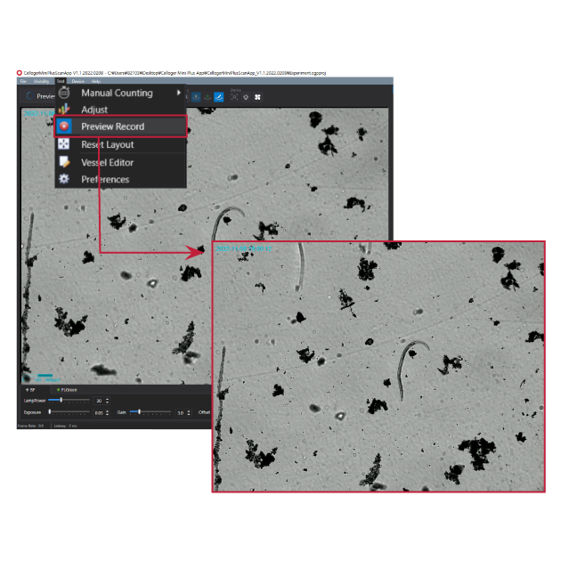
User-friendly functions
Autofocusing

Time-lapse video
Z-Stacking
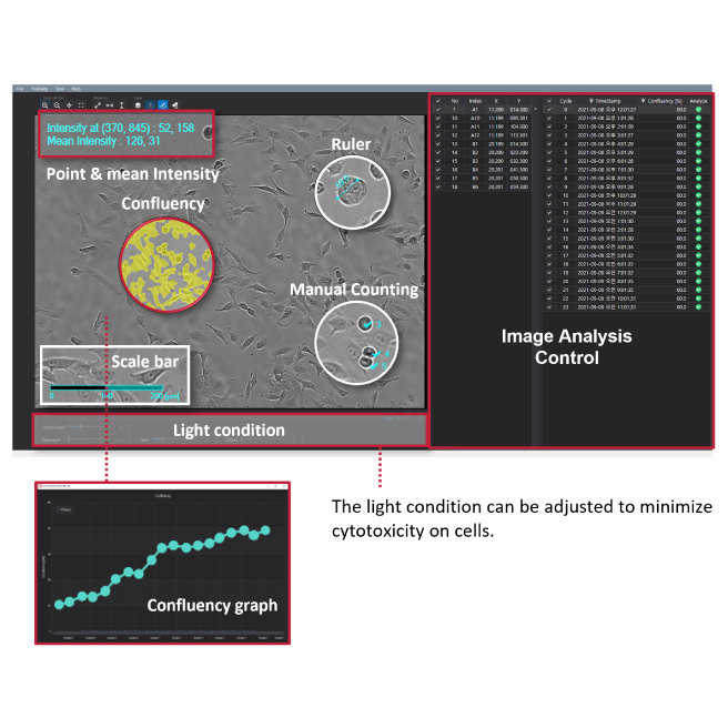
Analysis tools
Z-Stacking
Analysis tools

Specification
Ordering information
-
Application note_An integrated live cell monitoring_Celloger® Mini Plus_EN
2022.07.29
-
Application note_Analysis of Nocodazole-induced Cytotoxicity_Celloger® Mini Plus_EN
2022.10.26
-
Application note_Cellular assay using brightfield and fluorescence-based live cell imaging_Celloger® series
2023.11.28
-
Application note_Observation of Mitosis_Celloger® Mini Plus_EN
2022.09.22
-
Application note_Zebrafish observation using the Z stacking function_Celloger® Mini Plus_EN
2023.11.28

Celloger® Mini Plus I Cell proliferation
Observe the proliferation of HeLa cells in real-time every 1 hour for 110 hours.

Celloger® Mini Plus I Co-culture
Monitor the co-culture effects of K562 and NK92 cells (Calcein AM stained K562) for 18 hours.

Celloger® Mini Plus I Cytotoxicity
Cytotoxicity assay performed using time-lapse imaging of HeLa cells stained with green fluorescent dye.

Celloger® Mini Plus I Mitochondrial potential monitoring
The mitochondria of HeLa cells stained with TMRM were treated with CCCP to observe changes in fluorescence that indicate a crucial aspect of cellular health.

Celloger® Mini Plus I Neurite outgrowth
To observe neurite outgrowth in real-time, SH-SY5Y cells were stained with PKH26 dye on the third day of differentiation.

Celloger® Mini Plus I Nocodazole-mediated mitotic arrest
Time-lapse video of H2B-GFP transfected Hela cells treated with and without nocodazole.

Celloger® Mini Plus I Phagocytosis monitoring
Phagocytosis monitoring of LPS-stimulated Raw264.7 cell.
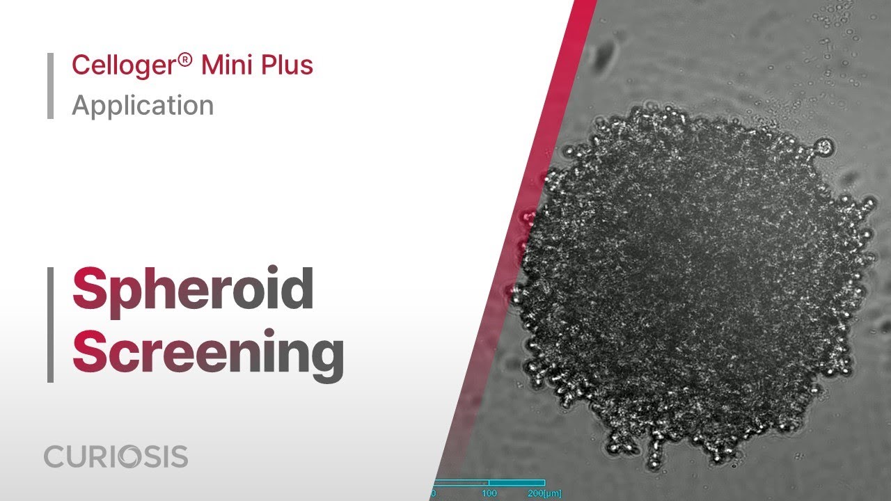
Celloger® Mini Plus I Spheroid screening
Monitoring concentration-dependent spheroid formation in two cell lines.

Celloger® Mini Plus I Wound healing assay
Wound healing assay was conducted using 4 different cell lines (HeLa, L929, CHO-k, U-2OS) and monitored at 30-minute intervals for 3 days.

Celloger® Mini Plus I Zebrafish observation
A transgenic Zebrafish (Larva) expressing green fluorescent protein was observed using the Celloger® Mini Plus Z-stacking function.